


Lateral Lumbar Interbody Fusion
This article focuses on just the surgical procedure of lateral lumbar interbody fusion. For a complete overview of spinal fusion, including approaches, bone grafting, complications, and rehabilitation, please go to Lumbar Spinal Fusion
Interbody Fusion
An interbody fusion is a method of fusing the lumbar spine that involves removing the intervertebral disk. When the disk space has been cleared out, a metal, plastic, or bone spacer is implanted between the two adjoining vertebrae.
These spacers, or "cages," usually contain bone graft material. This promotes bone healing and facilitates the fusion. After the cage is inserted, surgeons often use metal screws, plates, and rods to further stabilize the spine.
Lateral Lumbar Interbody Fusion
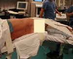
A patient in position for lateral lumbar interbody fusion.
An interbody fusion can be performed using a variety of different approaches. For example, a surgeon can access the spine through incisions in the lower back or through incisions in the front of the body.
In a lateral lumbar interbody fusion, the surgeon takes a side approach and centres the incision over the patient's flank. With this approach, the surgeon can reach the vertebrae and intervertebral disks without moving the nerves or opening muscles in the back.
The lateral approach is often referred to as extreme lateral or direct lateral interbody fusion (XLIF or DLIF).
Surgical Technique
During the surgery, the patient is placed in the side position and the table is bent to provide the surgeon with a maximum view of the spine.
In some cases, an instrument called a tubular retractor is inserted through the skin and soft tissues down to the spinal column. The tubular retractor holds the muscles open and gives the surgeon a clear view of the spine.
During the procedure, the disk is removed and a cage packed with bone graft is inserted between the vertebrae. Titanium screws are used to hold the cage in place. In some cases, an additional incision is made on the back to insert the screws.
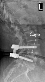
This x-ray taken from the side shows the cage between the vertebrae and screws that are stabilizing the spine.
Advantages of Lateral Lumbar Interbody Fusion
Each surgical approach — whether from the front, back, or side — has advantages and disadvantages. The possible advantages of a lateral lumbar fusion include:
- Less damage to the midline back muscles
- Easier access to the spine, in many cases
- Improved alignment of the spinal bones
In addition, a lateral fusion may be performed with a less invasive technique, resulting in reduced muscle injury.
These results can also be achieved through fusions performed from the back or the front. Talk to your surgeon about the approach that would best meet your health needs.