


Intervertebral Cages
Table of Contents
Rationale
The intervertebral fusion cage is another tool for the spine surgeon to use in helping treat various low back problems. For patients that require fusion surgery to treat degenerative disc disease, the intervertebral fusion cage has been shown to be effective for several reasons.
- Low complication rate
- Minimized pain after surgery due to less trauma during surgery
- Shorter hospital stay compared to other types of fusion methods
- Quicker return to daily activities
The cage is not designed to treat all types of spinal problems. You may be a candidate if your pain is from degenerative disc disease with segmental instability.
Surgical Devices
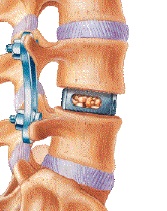
The intervertebral fusion cage is a hollow cylinder. The cages are made from various materials including metal or carbon graphite fibre. Doctors place bone graft inside the cylinder. The holes in the cage keep the graft in contact with the bony surface of the vertebrae. This ensures that the bone graft unites with the vertebrae, forming a solid fusion.
The cage helps in several ways. The solid cage separates and holds two vertebrae apart. This makes the opening around the nerve roots bigger, relieving pressure on the nerves. As the vertebrae separate, the ligaments tighten up, reducing instability and mechanical pain. The cage also replaces the problem disc while holding the two vertebrae in position until fusion occurs.
The intervertebral fusion cage is a new device. However, it has been studied in the U.S. for over ten years. There have been well over 10,000 procedures performed using intervertebral fusion cages, and it has proven to be a safe procedure. Several of the devices have been approved for use by the United States Food and Drug Administration (FDA).
Procedure
Cages can be implanted from the front or back of the spine. Surgery from the back of the spine may be needed to remove bone spurs or a herniated disc into the spinal canal. In these instances, the cages can be implanted from the back, without having to make an additional incision in the patient's abdomen.
Most often, the cages are put in during surgery through the front of the spine. Working from the front can be done with an open procedure, where a large incision is made through the abdomen. This procedure is being perfected with the use of a laparoscope, a TV camera that allows the doctor to see inside the abdominal cavity while working on the spine. This method only requires a few small incisions, which seems to help patients heal and get moving faster after surgery. Using a laparoscope for this procedure is difficult and may not be possible in all cases.
To see and work on the problem disc from the front, the surgeon makes an incision in the abdomen. The size of the incision depends on whether a laparoscope will be used. Organs and vessels are carefully moved aside. The problem disc is located using another type of special instrument called a fluoroscope (a special X-ray machine that shows the images on a TV screen).
In most cases, two cages are placed side by side to replace one disc. The surgeon drills two holes through front of the disc. Before putting one cage into each hole, the doctor prepares the cages. Bone graft may be taken from your pelvis bone through a small incision on your side. A second incision may not be needed if your surgeon uses a bone graft substitute.
The doctor packs the cages with the bone graft and inserts the cages side by side into the drill holes. A fluoroscope is used to make sure the cages are in the correct position. The abdominal incisions are sewn together, completing the surgery.