

Spinal Fusion Glossary: Spine Basics
Understanding why spinal fusion is done can be difficult and confusing. Your surgeon will discuss your questions in detail. To help you better understand the different spinal fusion surgeries, this glossary of words and abbreviations has been developed.
Spine Basics
Understanding your spine and how it works can help you better understand some of the problems that occur from aging or injury.
Many demands are placed on your spine. It holds up your head, shoulders, and upper body. It gives you support to stand up straight, and gives you flexibility to bend and twist. It also protects your spinal cord.
Spinal Curves
Your spine is made up of three segments. When viewed from the side, these segments form three natural curves. The "c-shaped" curves of the neck (cervical spine) and lower back (lumbar spine) are called lordosis. The "reverse c-shaped" curve of the chest (thoracic spine) is called kyphosis.
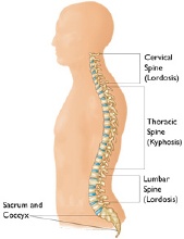
These curves are important to balance and they help us to stand upright. If any one of the curves becomes too large or small, it becomes difficult to stand up straight and our posture appears abnormal.
Abnormal curvatures of the spine are also referred to as spinal deformity. These types of conditions include kyphosis of the thoracic spine ("hunchback") and lordosis of the lumbar spine ("swayback").
Scoliosis is another type of spinal deformity. When viewing the spine from the front or back, scoliosis is a sideways curvature that makes the spine look more like an "S" or a "C" than a straight "I."
Parts of the Spine
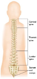
The spine from the back.
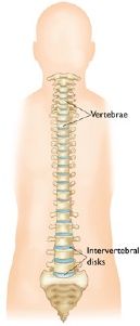
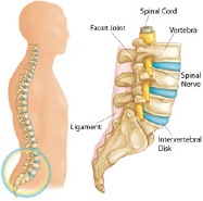
Your spine is made up of small bones, called vertebrae, which are stacked on top of one another and create the natural curves of your back.
Vertebrae
These bones connect to create a canal that protects the spinal cord.
Parts of the lumbar spine.
The cervical spine is made up of seven small vertebrae that begin at the base of the skull and end at the upper chest. The thoracic spine is made up of 12 vertebrae that start from the upper chest to the middle back and connect to the rib cage. The lumbar vertebra consists of five larger vertebrae. These vertebrae are larger because they carry more of your body's weight.
Spinal Cord and Nerves
The spinal cord extends from the skull to your lower back and travels through the middle part of each stacked vertebra, called the central canal. Nerves branch out from the spinal cord through openings in the vertebrae and carry messages between the brain and muscles.
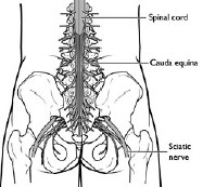
The cauda equina.
The spinal cord ends around the first and second lumbar vertebrae in the lower back and continues as nerve roots. This bundle of nerve roots is called the cauda equina. They exit the spinal canal through openings in the vertebrae (foramen), just like other nerve roots. In the pelvis, some of the nerves group into the sciatic nerve, which extends down the leg.
Muscles and Ligaments
These provide support and stability for your spine and upper body. Strong ligaments connect your vertebrae and help keep the spinal column in position.
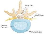
Healthy intervertebral disk (cross-section view).
Intervertebral Disks
Intervertebral disks sit in between the vertebrae. They are flat and round, and about 1.3 cm thick.
Intervertebral disks are made up of two components:
- Nucleus pulposus. The nucleus pulposus is jelly-like and makes up the centre of the disk. The jelly is partly made of water and gives the disk flexibility and strength.
- Annulus fibrosus. This is the flexible outer ring of the disk. It is made up of several layers, like elastic bands.
When you are standing, or moving, weight is put onto the nucleus. In response, the nucleus expands. The annulus holds the nucleus in place. This allows movement to take place, yet maintains the strength of the spine. In effect, disks act as shock absorbers for the spine.
The intervertebral disk is a very important structure. Many nerve endings supply the annulus and, as a result, an injured annulus can cause pain.
Facet Joints
Between the back of the vertebrae are small joints that also help your spine move. These facet joints have a cartilage surface, very much like a hip or a knee joint does. The facet joints are important for allowing rotation of the spine but may develop arthritis and become a source for low back or neck pain.
Incision Site
Surgeons can reach the spine by making an incision (cut) in different places on your body. Incision sites are often described as:
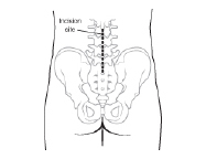
A posterior incision site.
Anterior. This term refers to the front of your body. In spinal fusion surgery, an anterior fusion is done by making an incision in the abdomen (belly).
Posterior. This refers to the back part of your body. If you are having a posterior fusion in your lower back, you will lie on your belly during the operation and your surgeon will make the incision in your lower back.
Lateral. This refers to the side part of your body. Surgeons can reach certain parts of the lumbar spine by making an incision in your side.
Procedure Site
Surgeons will also perform the surgical procedure in specific parts of your spine, such as the vertebra or the intervertebral disk. In most cases, the part of your spine being operated on is the place where the problem is present.
Inter-transverse or posterolateral. This is the part of the lumbar spine between the transverse processes of a vertebra.
Surgeons most often reach this area by making an incision on the back. This type of fusion is called:
- Posterior (from the back) posterolateral (specific spinal anatomy involved) F This procedure is often referred to as a PLF.
Interbody. This is the part of the spine where the disk is present, between the bones (vertebrae). An interbody fusion can be performed with different approaches:
- Anterior (from the belly) Lumbar (part of the spine being operated upon) Interbody (in the disk space) F You may see this written as an ALIF.
- Posterior (from the back) Lumbar (part of the spine being operated upon) Interbody (in the disk space) F or PLIF.
- A different type of PLIF is called Although the spine is still reached through an incision in the back, the disk space is approached from the side. Transforaminal (from the back) Lumbar (part of the spine being operated upon) Interbody (in the disk space) Fusion or TLIF.
- Lateral (from the side) Lumbar (part of the spine being operated upon) Interbody (in the disk space) Fusion (specific surgery being done). This type of surgery is often described as a Direct lateral or Extreme lateral approach (DLIF or XLIF).